What is the human blastocyst?
By Galia Tsangkalova – Embryologist, Lab Director
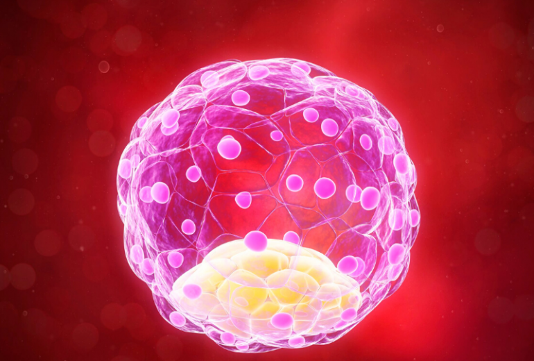
Human blastocyst is an embryo on its 5th day of development. It consists of 24 up to 322 cells, its size is about 0.2mm and its discrete parts are: the trophectoderm (TE), the inner cell mass (ICM) and the blastocoel. All these structures are surrounded by zona pellucida (ZP), a thick glycoprotein protecting coat that is present from the oocyte to the blastocyst stage. The side of the blastocyst where the ICM is formed is called the animal pole and the opposite side is the vegetal pole.

- TE is the outer part of the embryo, a sphere like layer of cells which will form mostly extra embryonic tissues as the placenta.
- ICM is the inner part of the embryo, a cluster of cells, attached on the wall of the TE layer, which will go on to form the actual fetus.
- Blastocoel is a cavity filled with fluid which defines the blastocyst expansion. The fluid is produced by the TE cells.
The initial TE and ICM cells are present on the pre-blastocyst stage, called morula (4th day of embryo development), when no cavitation is observed. A defining moment between Day 4 and 5 is when TE cells start to secrete fluid and as they divide, the fluid accumulates so the cavity is gradually formed. The newly formed blastocyst on day 5 starts to enlarge due to the increase of the TE cell number, the volume of the blastocoels and the pressure inside the cavity. Alongside, the consequent progressive thinning of the ZP, results to the hatching procedure that begins with the formation of a small hole on ZP. On Day 5 or usually on Day 6, the pressure rises inside the blastocyst, the hole expands, and finally the whole blastocyst comes out of its shell, fact that allows increased growth, access to uterine nutrient secretions and blastocyst adhesion to the uterine lining. Once the hatching takes place, the blastocyst is ready for implantation around Day 7.
What is the human blastocyst?

By Galia Tsangkalova – Embryologist, Lab Director
Human blastocyst is an embryo on its 5th day of development. It consists of 24 up to 322 cells, its size is about 0.2mm and its discrete parts are: the trophectoderm (TE), the inner cell mass (ICM) and the blastocoel. All these structures are surrounded by zona pellucida (ZP), a thick glycoprotein protecting coat that is present from the oocyte to the blastocyst stage. The side of the blastocyst where the ICM is formed is called the animal pole and the opposite side is the vegetal pole.

- TE is the outer part of the embryo, a sphere like layer of cells which will form mostly extra embryonic tissues as the placenta.
- ICM is the inner part of the embryo, a cluster of cells, attached on the wall of the TE layer, which will go on to form the actual fetus.
- Blastocoel is a cavity filled with fluid which defines the blastocyst expansion. The fluid is produced by the TE cells.
The initial TE and ICM cells are present on the pre-blastocyst stage, called morula (4th day of embryo development), when no cavitation is observed. A defining moment between Day 4 and 5 is when TE cells start to secrete fluid and as they divide, the fluid accumulates so the cavity is gradually formed. The newly formed blastocyst on day 5 starts to enlarge due to the increase of the TE cell number, the volume of the blastocoels and the pressure inside the cavity. Alongside, the consequent progressive thinning of the ZP, results to the hatching procedure that begins with the formation of a small hole on ZP. On Day 5 or usually on Day 6, the pressure rises inside the blastocyst, the hole expands, and finally the whole blastocyst comes out of its shell, fact that allows increased growth, access to uterine nutrient secretions and blastocyst adhesion to the uterine lining. Once the hatching takes place, the blastocyst is ready for implantation around Day 7.


